Thrives Article

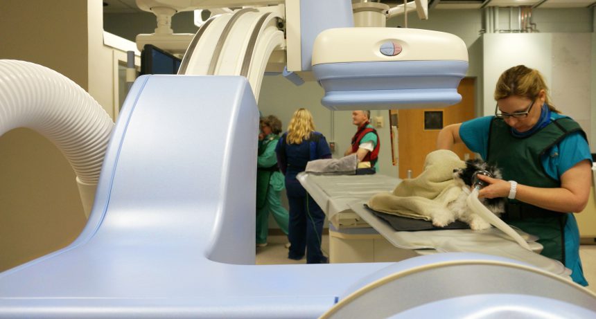 Veterinary Diagnostic Imaging Veterinary diagnostic imaging is an important part of a pet’s medical treatment plan. X-rays are used to help diagnose broken bones, foreign objects in the digestive tract and other common problems. Digital x-rays reduce the pet’s exposure to radiation and allows to see more detail. Ultrasounds are also an excellent diagnostic tool. They allow us to see internal organs and measure blood flow and other parameters.
Magnetic resonance imaging (MRI) is an advanced diagnostic technique that uses a powerful magnet to create three-dimensional representations of structures within the body. It is the best test to evaluate tumors, abscesses and changes in blood vessels. MRI is also very useful in evaluating spinal cord injuries and syringomyelia. Computed tomography (CT) is a computer-enhanced x-ray procedure that generates cross-sectional images of the body. It is often combined with a nuclear medicine scan to get additional information about how an organ is functioning. Nuclear medicine involves injecting a short-acting radioactive dye and then using a gamma camera to detect the emission of radiation. It is used to assess liver, thyroid, and kidney function as well as bone disease. Veterinary radiology is a highly specialized discipline that requires expertise to interpret images and provide the appropriate care for the pet. Despite the importance of Veterinary Diagnostic Imaging, it is not without its limitations. Not all fractures are detected by x-rays, results of imaging suggest that a fracture is present when none is actually present and/or results of imaging result in incorrect clinical management decisions. Veterinary diagnostic imaging techniques are becoming common in veterinary practice and research. X-rays are the most commonly used type of veterinary diagnostic imaging. However, they may not reveal certain conditions. An ultrasound uses sound waves to examine internal organs, and it is usually non-invasive and non-painful. Veterinary CT is a fast and efficient imaging modality that provides a wealth of information to the clinician. It is a valuable diagnostic tool that can evaluate many soft tissue structures and diseases traditionally diagnosed by radiography such as gastrointestinal and respiratory tract disease and can also be used to aid in surgical planning, radiation therapy treatment and for assessing bone health. Much like traditional radiographs, CT scans require an animal to be in a very still position and therefore most studies are performed under general anesthesia. This is important to ensure that the resulting images are of good quality and do not reflect movement in a manner similar to ‘motion artefact’. Most veterinary CT studies are contrast enhanced using nonionic iodinated contrast media which allows for greater differentiation of structures on the image and is particularly useful in evaluating vascular disease. This is a common imaging modality in human medicine and is gaining traction in veterinary practice as well. MRI produces high-contrast, anatomically detailed tomographic images that don’t reflect tissue density like radiography. However, unlike radiography and CT, MRI does not use ionizing radiation. Increasingly, veterinary clinics and hospitals are investing in their own refurbished MRI machines. They are especially useful for diagnosing conditions that cannot be assessed using other imaging techniques such as those involving the brain and spinal cord. Nuclear scintigraphy is a diagnostic imaging technique that allows veterinarians to see structural changes in horse’s bones and soft tissues before they can be seen on radiographs. It involves injecting a small amount of a radioactive substance which circulates throughout the body and localizes to areas with increased bone turnover or blood supply. When this radiation is detected by a special device called a gamma camera, it produces images that radiologists use to diagnose problems. Ultrasonography is a painless diagnostic imaging technique that uses digital sound waves to produce an image of the pet’s internal structures. This technology has a wide variety of applications in veterinary medicine including scanning abdominal organs, cardiac chambers and valves, the liver and gallbladder, and reproductive organs.
0 Comments
Leave a Reply. |
